Bone Cancer: A Comprehensive Overview and Advanced Detection Techniques
Bone cancer, a formidable adversary in the realm of human health, originates from the cells residing within our skeletal framework. This insidious disease manifests in two distinct forms: primary and secondary. Primary bone cancer, a self-contained entity, arises from the bone cells themselves, while its secondary counterpart originates elsewhere in the body and subsequently infiltrates the bone tissue. Early detection and prompt intervention are paramount in combating bone cancer, as they can significantly improve patient outcomes and prevent the relentless spread of cancerous cells to other body regions. This comprehensive analysis delves into the intricacies of bone cancer, exploring its classification, symptoms, diagnosis, and the cutting-edge detection techniques that are revolutionizing the fight against this formidable foe.
Classification of Bone Cancer: Unveiling the Two Main Types
Bone cancer, in its diverse manifestations, can be broadly categorized into two primary types:
1. Primary Bone Cancer: This type emerges from the cells within the bone itself, instigating a localized battle against healthy tissue.
2. Secondary Bone Cancer: This type, a cunning infiltrator, originates from a distant site within the body, stealthily spreading its malignant tentacles to the bone, where it wreaks havoc upon the unsuspecting cells.
Symptoms of Bone Cancer: Recognizing the Warning Signs
In its insidious early stages, bone cancer may lurk silently, revealing no telltale signs of its presence. However, as the disease progresses, it often manifests a range of symptoms that demand attention:
– Persistent bone pain, a nagging reminder of the cancer’s relentless assault.
– Swelling or tenderness near the affected area, a visible manifestation of the underlying struggle.
– Difficulty in movement, a hindrance to everyday activities, caused by the disruptive presence of cancer.
– Unexplained weight loss, a consequence of the body’s diverted resources and metabolic imbalances.
– Fatigue, a debilitating weakness that saps the patient’s energy and zest for life.
– Night sweats, a discomforting disturbance during the hours of slumber, triggered by the body’s internal turmoil.
– Fractures with minimal or no trauma, a sign of the bone’s weakened structure, succumbing to the cancer’s insidious weakening.
Diagnosis of Bone Cancer: Unraveling the Enigma
Proper diagnosis of bone cancer entails a meticulous investigation, employing a combination of patient history, physical examination, and advanced imaging techniques. This multi-pronged approach aims to unveil the hidden secrets of the disease, guiding the path towards appropriate treatment.
– X-ray: The initial foray into the realm of bone cancer diagnosis, providing rudimentary insights into bone abnormalities.
– Computed Tomography (CT): A more sophisticated imaging modality, offering detailed cross-sectional images of bones and surrounding tissues, revealing intricate details of the cancerous invasion.
– Magnetic Resonance Imaging (MRI): A non-invasive journey into the depths of the body, generating detailed images of bones and soft tissues, aiding in the assessment of tumor extent and involvement of surrounding structures.
– Positron Emission Tomography (PET): A metabolic sleuth, identifying areas of heightened activity within the bone, assisting in tumor detection and staging, guiding the treatment strategy.
Role of Medical Imaging: A Beacon in the Darkness
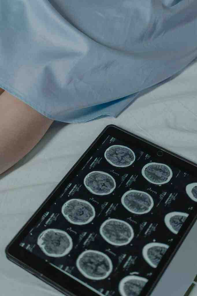
Medical imaging, a beacon of hope in the fight against bone cancer, plays a pivotal role in its early detection, enabling timely intervention and improved patient outcomes. Radiologists, the guardians of medical images, often prefer these techniques due to their advantages in time management, cost-effectiveness, and the invaluable ability to facilitate early detection, potentially altering the course of the disease.
Image Processing and Machine Learning: Unveiling Hidden Patterns
Recent advancements in image processing and machine learning techniques have heralded a new era in bone cancer detection, offering innovative approaches that empower healthcare professionals in their quest to combat this formidable disease. These techniques, like skilled detectives, meticulously analyze medical images, uncovering hidden patterns and abnormalities that may escape the human eye.
– Preprocessing: The initial step, akin to preparing a crime scene for investigation, involves removing noise and artifacts from images, enhancing their quality for further analysis, ensuring clarity for accurate interpretation.
– Segmentation: The process of dividing the image into regions of interest, isolating the cancerous area from healthy tissues, akin to separating the wheat from the chaff, enabling focused analysis of the affected region.
– Feature Extraction: The identification and extraction of relevant features from the segmented images, akin to collecting evidence at a crime scene, aiding in the classification process, providing crucial clues for diagnosis.
– Classification: The final verdict, utilizing machine learning algorithms to classify images as cancerous or non-cancerous based on the extracted features, akin to a jury delivering its judgment, guiding the path towards appropriate treatment.
Deep Learning: A Revolutionary Force in Bone Cancer Detection
Deep learning, a subset of machine learning, has emerged as a formidable ally in the fight against bone cancer, demonstrating remarkable accuracy in classifying bone cancer images. Deep learning models, particularly Convolutional Neural Networks (CNNs), possess an uncanny ability to learn complex patterns and relationships within medical images, mimicking the keen eye of an experienced radiologist.
Current Study: Illuminating the Path to Early Detection
This groundbreaking study focuses on the early detection of three prevalent bone cancers: parosteal osteosarcoma, enchondroma, and osteochondroma, utilizing a comprehensive dataset of CT images. The study employs a synergistic combination of image processing techniques and deep learning models to achieve accurate cancer detection, potentially revolutionizing the diagnosis and treatment of bone cancer.
Methodology: A Step-by-Step Approach
1. Data Collection: The foundation of the study, assembling a vast dataset of 1141 bone CT images, encompassing both normal and cancerous cases, ensuring a comprehensive representation of the disease.
2. Preprocessing: Preparing the images for analysis, removing noise and artifacts, akin to decluttering a room, enhancing image quality for subsequent analysis, ensuring clarity and accuracy.
3. Segmentation: Isolating the cancerous regions from healthy tissues, akin to separating the wheat from the chaff, employing K-means clustering and Canny edge detection techniques, providing focused analysis of the affected areas.
4. Feature Extraction: Identifying and extracting relevant features from the segmented images, akin to collecting evidence at a crime scene, employing statistical and texture analysis techniques, providing crucial clues for classification.
5. Classification: The final verdict, utilizing CNN models to classify images as cancerous or non-cancerous based on the extracted features, akin to a jury delivering its judgment, guiding the path towards appropriate treatment.
Results and Discussion: Unveiling the Findings
The study yielded promising results in bone cancer detection using CT images, demonstrating the immense potential of image processing and deep learning techniques in improving patient outcomes. The CNN models exhibited remarkable accuracy in classifying normal and cancerous images, indicating their potential for clinical applications. These findings suggest that the proposed methodology can empower radiologists in detecting bone cancer at an early stage, potentially altering the course of the disease and improving patient survival rates.
Conclusion: A Glimmer of Hope in the Fight Against Bone Cancer
This study stands as a testament to the effectiveness of image processing and deep learning techniques in bone cancer detection using CT images. The proposed methodology provides a valuable tool for radiologists, assisting them in the early detection of bone cancer and facilitating timely intervention. Future research endeavors will delve deeper into the realm of possibilities, exploring the use of larger datasets, investigating the application of other deep learning models, and integrating clinical data to further enhance the accuracy and reliability of bone cancer detection. These advancements hold the promise of revolutionizing the fight against bone cancer, offering hope and improved outcomes for patients battling this formidable disease.
